
Digital analysis of radiograms in traumatology and orthopedics
DIGITAL ANALYSIS OF RADIOGRAMS IN TRAUMATOLOGY AND ORTHOPEDICS
The tasks of digital analysis of radiographs:
• Achieving optimal visibility of the picture
• Obtaining objective quantitative and qualitative data on the density and structure of bone tissue
Object of analysis:
• Analog images
• Digital imaging
The purpose of the study:
• Possibility to observe the dynamics of changes in the studied parameters
• Document the results of the study
• Creation of illustrative material for scientific publications, dissertation research
METHODS OF DIGITAL ANALYSIS OF RADIOGRAMS IN TRAUMATOLOGY AND ORTHOPEDICS:
• Optical densitometry
• Color contrasting
• Building optical relief
• Combination of methods
• Graphic image analysis
COLOR CONTRASTING OF RADIOGRAPHS
The essence of the color contrasting method consists in converting a black-and-white image of a radiograph into a color image based on features that reflect certain properties of the image, in this case, its optical density (brightness).
The advantage of using color contrasting lies in the creation of an additional information feature that makes it possible to objectify the process of interpreting an image due to the fact that the human eye distinguishes more colors than shades of any one color, which occurs on a radiograph.
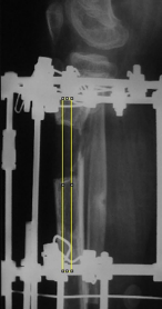
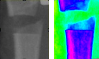 |
| Color contrast radiography of the lower leg of a patient with achondroplasia two weeks after limb lengthening surgery |
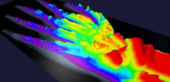
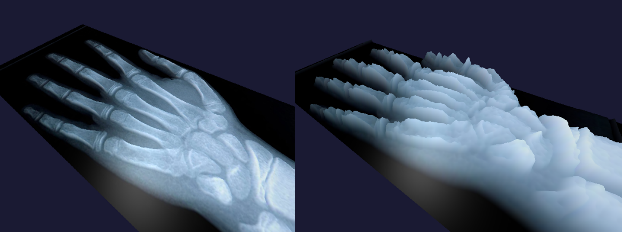
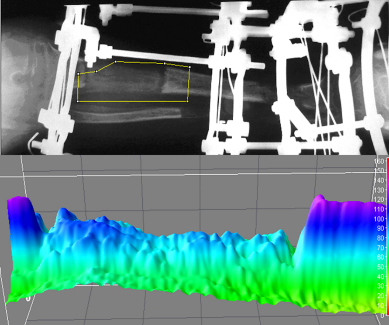
Construction of the optical relief of the X-ray image
The essence of the method for constructing the optical relief of an x-ray image is to obtain a 3D image of a relief surface in which the height of each section is a function of the brightness (density) of the original image at a given point, in parallel, it is possible to obtain quantitative data on the selected fragments with their subsequent processing by methods of mathematical statistics.
 |
|
| X-ray of the hand on a 3D surface | 3D surface (relief) of radiographs of the hand in 3D space |
Combining the method of color contrasting and the method of constructing an optical relief
 |
| Graphical image analysis: profilogram |
THE METHOD FOR CONSTRUCTING AN IMAGE PROFILOGRAM CONSISTS IN CONSTRUCTING A LINE GRAPH REFLECTING THE CHANGE IN IMAGE BRIGHTNESS ALONG A LINE THAT IS ARBITRARILY SELECTED ON THE STUDIED PART OF THE IMAGE
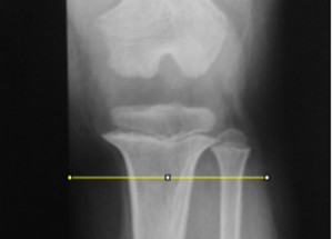 |
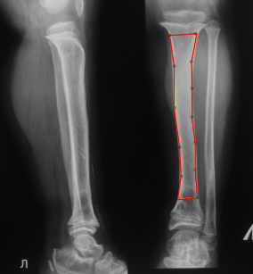
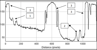
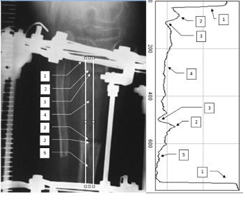
| Line for constructing a profilogram of the tibia |
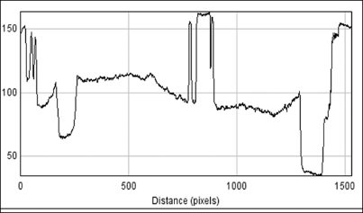
Graphical image analysis: profilogram
 |
| Graphical image analysis: profilogram
1 – tibia, 2 – cortical plates, 3 – cavity of the medullary canal |
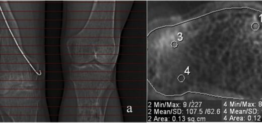
QUANTITATIVE DATA OF STUDY RESULTS AND THE MEANS OF THEIR REPRESENTATION
Mathematical methods of statistical analysis:
- mode
- median
- average value of image brightness
- variance
- coefficient of variation of a statistical parameter
Graphical methods for presenting statistical indicators:
- tables
- Histograms
- Graphs
- Diagrams
 |
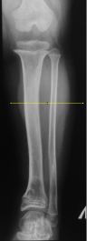
| X-ray of the lower leg of a patient with achondroplasia and the study area |
Statistical indicators of the results obtained
 |
| Distribution histogram and statistical indicators of the results obtained
(number of indicators, average value of the indicator, standard deviation of the minimum and maximum values, the mode of the indicator and its volume) |
Graphical analysis of study results in dynamics
 |
 |
| Before surgery
On the profilogram of the tibia, there are no two peaks of elevation characteristic of the cortical plates, which clearly indicates the absence of the medullary canal. However, the tibial plateau, curved downwards, indicates the process of its formation. |
|
After operation
 |
 |
| On the profilogram of the tibia after bilocal, distraction osteosynthesis, it is shown
1 – diastasis between bone fragments 2 – compaction of the bone after its osteotomy 3 – details of the Ilizarov apparatus |
|
10 days after surgery
 |
 |
| Tibia profilogram after bilocal, distraction osteosynthesis 10 days of distraction | |
45 days after surgery
 |
 |
| The profilogram shows an increase in the density of the edges of the maternal bone directly adjacent to the regenerate (1). Optical profile of bone regenerate (2) | |
An example of a statistical analysis of the dynamics of reparative osteogenesis of a long tubular bone based on a digital analysis of the optical density of the bone renenerate image
 |
| From the above graph it follows that in the process of formation of the distraction regenerate, the ratio of structures of different density changes in a mirror way, which can serve as an indicator of the staging of the process and an indication for correcting the rate of distraction or removing the apparatus |
An example of a statistical analysis of the dynamics of the level of bone mineralization and regenerate based on a digital analysis of the optical density of an x-ray image
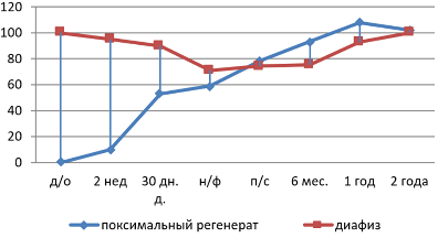 |
| It follows from the above graph that at the initial stage of bone lengthening, its osteoporosis develops, which is characterized by a decrease in the optical density of its image and the absence of minerals in the distraction regenerate. The middle stage is characterized by the absence of a medullary canal at the level of elongation and, as a result, an increase in the optical density of the regenerate, which exceeds the density of the bone. At the final stage, after the completion of the restructuring processes, the optical density of the bone is aligned along its entire length. |
VARIANTS OF THE COURSE OF REPARATIVE OSTEOGENESIS
Types of reparative osteogenesis
• Hyperplastic type
• Hypoplastic type
• Normotypic type
HYPERPLASTIC TYPE FORMATION OF DISTRACTION REGENERATE
Predisposing factors
• Multifragmented nature of the osteotomy
• Hypersthenic body type in a patient
• Features of the individual reaction of the body to skeletal injury
• Young age of the patient
• Multifragmentary fracture
• Discrepancy between the rate of distraction and the activity of reparative osteogenesis
• Preservation of the integrity of the periosteum during osteotomy or its detachment over a considerable extent with the organization of a hematoma under its surface.
Clinical and radiological signs of the hyperplastic form of the regenerate
• Violent periosteal reaction
• The transverse dimensions of the regenerate exceed the diameter of the maternal bone
• There is also an increased optical density of the regnerate in comparison with the average value of this indicator
• The structure of the regenerate is homogeneous
Hyperplastic type of distraction regenerate formation
 |
| Hyperplastic type of distraction regenerate formation |
HYPOPLASTIC TYPE FORMATION OF DISTRACTION REGENERATE
Predisposing factors
• Patient with a slender physique
• Asthenic type of fusion
• Malnutrition
• Chronic diseases
• Non-pathic and dystrophic processes in the elongated limb
• Systemic diseases
• Features of the individual reaction of the body to skeletal injury
Clinical and radiological signs of the hypoplastic form of the regenerate
• Monotonous X-ray picture at repeated research.
• Heterogeneous structure of the regenerate
• The presence of areas of narrowing of the regenerate
• The optical density of the regenerate does not correspond to the terms and stage of treatment
 |
| Hypoplastic type of distraction regenerate formation |
SIGNS OF FORMATION OF DYSTROPHIC PHENOMENA IN THE HYPOPLASTIC DISTRACTION REGENERATE
Signs of the formation of dystrophic phenomena in the distraction regenerate
• Emergence of end plates
• The appearance of a resorption zone at the border with the maternal bone
• Distraction regenerate becomes sharply structured
• Its optical density remains the same or decreases.
• The ratio of mineralized structures is in sharp contrast
• The optical structure of the regenerate is sharply inhomogeneous and contains both structures of low density and structures of a high degree of mineralization
 |
| 1. details of the Ilizarov apparatus
2. end plate on the bone 3. zone of resorption of the regenerate 4. body regenerate 5. maternal bone |
Optical relief and color contrasting of the X-ray picture of the lower leg during the formation of dystrophic phenomena in the distraction regenerate
 |
| Optical relief and color contrasting of the roentgenogram of the lower leg during the formation of dystrophic phenomena in the distraction regenerate |
THE MAIN VALUE OF THIS STUDY
• Ability to compare indicators in dynamics
• Ability to obtain parametric indicators
• Possibility to document the research result
• Ability to present the received data in the form of tables, graphs, charts
• Ability to create illustrative material for scientific publications and dissertation research.
| Doctor of medical sciences, traumatologist-orthopedist of the traumatology and orthopedic department No. 14 of the Federal State Budgetary Institution “Research Center named after Academician G.A. Ilizarov “Ministry of Health of Russia, Kurgan |
| Konstantin I. Novikov – M.D, Ph.D., doctor traumatologist-orthopedist traumatology and orthopedic department №14, Federal State Budgetary Institution Scientific Research Center named after academician G.A. Ilizarova »Ministry of Health of Russia, Kurgan, e-mail: kinovikov@mail.ru |

What is the diagnostic potential of using this technique in clinical practice?
Big….:-)